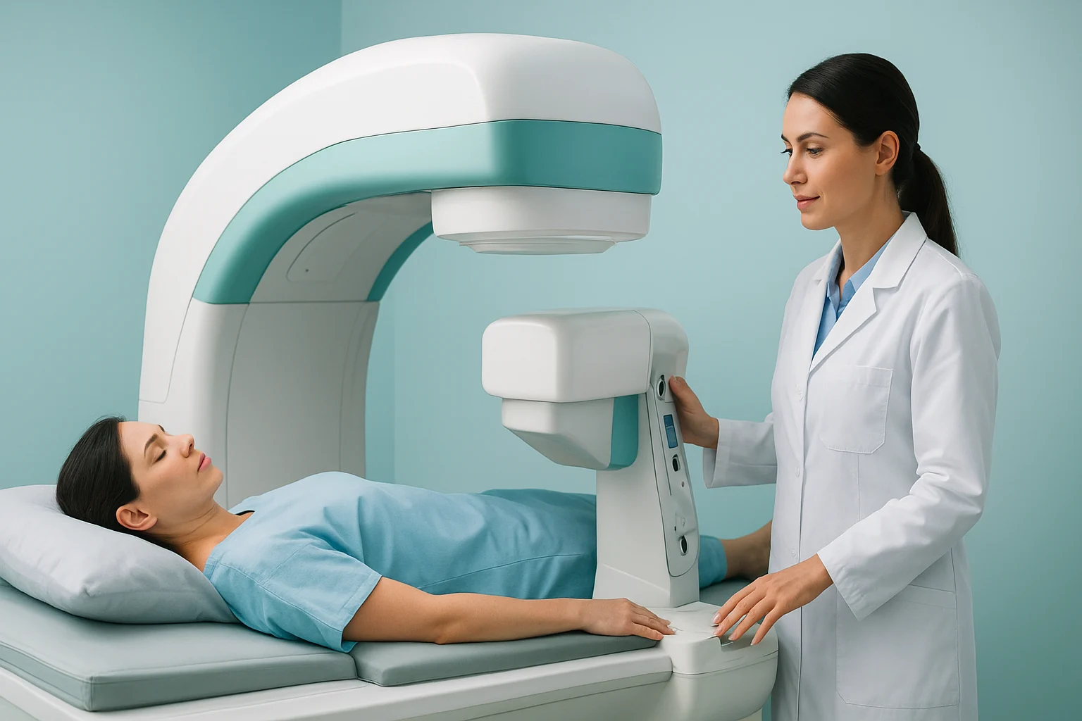

Bone scans are a valuable diagnostic tool when joint pain remains unexplained, even after an X-ray, MRI, or CT scan. They help doctors identify issues such as infection, implant loosening, or bone stress that may not be visible through standard imaging. This article explains how bone scans work, when they’re recommended, and what to expect from the procedure — so you can better understand your joint health journey.
A bone scan is a type of nuclear medicine imaging test that helps detect changes in bone metabolism. Unlike X-rays, which capture structural images, bone scans highlight areas of increased bone activity — often a sign that something is happening at the cellular level, such as infection, stress, or inflammation.
During the procedure, a small amount of radioactive tracer is injected into a vein. This tracer travels through the bloodstream and collects in the bones, especially in areas that are healing, infected, or inflamed. A special gamma camera then takes images that reveal patterns of tracer uptake, helping doctors pinpoint abnormalities invisible on other scans.

Bone scans are not always the first test used to investigate joint pain. Usually, doctors start with X-rays or MRI scans. However, when pain persists or symptoms don’t align with other imaging results, a bone scan can provide deeper insight.
Here are the most common reasons your doctor might recommend one:
When infection is suspected in a joint or bone, a bone scan can help confirm its presence and pinpoint the location. Infections cause increased blood flow and bone turnover, leading to a “hot spot” — an area of higher tracer uptake visible on the scan.
This is especially important when:
By identifying infection early, doctors can prescribe antibiotics or plan surgical treatment before the condition worsens.
Bone scans are also essential for assessing complications after joint replacement surgery. Over time, an artificial joint (such as a hip or knee implant) can become loose due to wear, bone resorption, or infection.
Typical signs include:
A bone scan helps distinguish between mechanical loosening (when the implant separates from the bone) and infection-related loosening (when infection weakens the connection). Sometimes, your doctor may request a specialized three-phase bone scan or SPECT/CT scan to provide more detailed functional and structural information.
If you’re experiencing persistent joint pain but standard imaging hasn’t revealed a cause, a bone scan can uncover subtle or hidden abnormalities. Examples include:
Bone scans are particularly valuable for detecting small lesions or changes before they become visible on X-rays, making them ideal for early diagnosis and intervention.
In some cases, bone scans are ordered to check whether a tumor or cancer has spread to the bones. These scans provide a comprehensive overview of skeletal activity, helping detect metastases or suspicious growths early.
While not a first-line test for joint pain, this use is vital when pain patterns suggest a more serious underlying condition.
If your doctor recommends a bone scan, here’s what typically happens:
The test is non-invasive, and side effects are extremely rare. You can resume normal activities right after.
Bone scans provide unique advantages in musculoskeletal diagnosis:
Early Detection:
Identifies subtle changes in bone metabolism before structural damage appears on X-ray or MRI.
Whole-Skeleton Overview:
Evaluates multiple joints at once to detect systemic or widespread bone disorders.
Differentiation:
Helps distinguish between infection, loosening, and inflammation — conditions that may look similar on other scans.
Guided Treatment:
Directs physicians toward the most appropriate management plan, including surgery, antibiotics, or physiotherapy.
Limitations of Bone Scans
While bone scans are powerful diagnostic tools, they do have some limitations:
They can’t always identify the exact cause of increased activity (additional tests may be needed).
They involve a small amount of radiation exposure — though less than a CT scan.
Some conditions, like arthritis or fractures, may produce similar scan results.
Because of this, bone scans are often used in combination with other imaging techniques or laboratory tests for a clearer picture.
Once your results are available, your doctor will review them alongside your symptoms and medical history.
For example:
Your orthopaedic specialist will interpret these findings to determine the best course of treatment.
1. Are bone scans safe?
Yes. Bone scans use a very small amount of radioactive material, which leaves your body naturally through urine within 24 hours. The radiation dose is minimal and considered safe for most patients.
2. How long does a bone scan take?
The entire process takes about 3–4 hours, including waiting time for the tracer to circulate. The actual scanning usually takes 30–60 minutes.
3. Can bone scans detect arthritis?
Yes. They can show early inflammation and bone remodeling associated with arthritis — even before it’s visible on X-rays.
4. What’s the difference between a bone scan and an MRI?
An MRI shows detailed images of soft tissues and bone structure, while a bone scan highlights bone activity and metabolic changes. The two are often complementary.
5. How should I prepare for a bone scan?
You don’t need to fast. Drink water before and after the test to help eliminate the tracer. Be sure to tell your doctor if you’re pregnant or breastfeeding.
6. Can bone scans show joint implant problems?
Absolutely. Bone scans are one of the best ways to detect implant loosening or infection after a hip or knee replacement.
Bone scans play a crucial role in uncovering hidden causes of joint pain, especially when other tests fall short. From detecting infection and implant loosening to explaining persistent pain, they provide valuable information that guides accurate diagnosis and treatment.
If you’re dealing with ongoing joint pain, your orthopaedic specialist — such as Dr Oliver Khoo — can determine whether a bone scan is right for you.
For related resources on joint health and orthopaedic care, visit the Oliver Khoo Blog