Dr Oliver Khoo
Dr Oliver Khoo is a leading orthopaedic surgeon based in Australia, specialising in hip, knee, and shoulder joint replacement, revision surgery, and complex fracture care. With years of experience and a patient-centred approach, Dr Khoo delivers both excellence in the operating theatre and clarity in patient education.
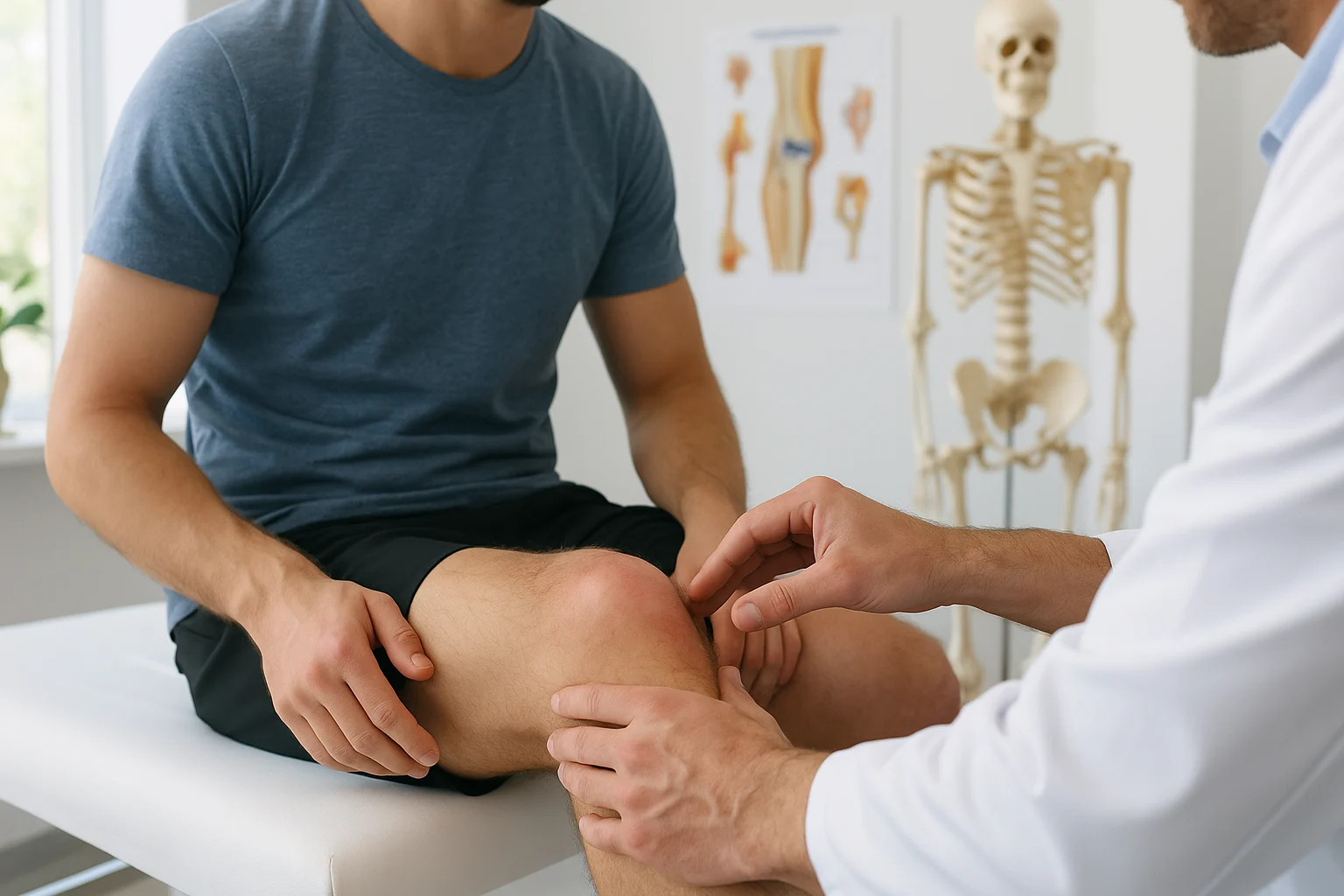
1. Why Accurate Assessment Matters
Knee injuries are among the most common musculoskeletal issues seen in both general practice and emergency settings. Whether it’s a sports-related incident, a workplace accident, or a simple household fall, identifying the exact nature of the injury early on is crucial.
Failing to assess thoroughly can lead to:
- Missed ligament injuries, such as ACL or PCL tears, which may result in persistent knee instability.
- Undiagnosed meniscal tears, potentially causing mechanical symptoms like locking and long-term degeneration.
- Unnecessary imaging, increasing healthcare costs and fuelling patient anxiety without adding clinical value.
Understanding the injury begins with a solid history and physical exam, followed by the judicious use of imaging where indicated.
2. History & Examination: The Cornerstones of Diagnosis
Mechanism of Injury – What Happened Matters
Carefully listening to how the injury occurred can reveal much about the underlying damage.
- Twisting with pivoting or deceleration → often implicates the ACL or meniscus.
- Direct contact to the lateral or medial knee → think MCL, LCL, or bone bruise/fracture.
- Hyperextension or landing awkwardly from height → raise suspicion for PCL injury or even posterior capsule strain.
Symptom Timing – Clues from the Swelling
- Immediate swelling (within 2 hours) usually indicates haemarthrosis, commonly due to an ACL rupture, patellar dislocation, or intra-articular fracture.
- Delayed swelling (6–12 hours) is more often seen with meniscal injuries or capsular sprains.
- Locking, catching, or clicking are red flags for meniscal tears, especially when associated with joint line tenderness.
3. Ottawa Knee Rules – When to X‑Ray
Not every knee injury needs imaging on the first presentation. In fact, most soft tissue injuries won't show up on a plain X-ray. That’s where the Ottawa Knee Rules come in—they help clinicians safely rule out fractures without overusing radiographs.
You should order a knee X-ray only if any of the following are present:
- Age 55 years or older
- Isolated tenderness over the patella (with no other bone tenderness)
- Tenderness at the head of the fibula
- Inability to flex the knee to 90 degrees
- Inability to bear weight both immediately after injury and in the emergency department (defined as taking four steps independently, regardless of limp)
If none of these are present, the likelihood of a significant fracture is less than 1%—making imaging unnecessary in most low-risk cases.
Why does this matter?
- It avoids exposing patients to unnecessary radiation
- Reduces healthcare costs and delays
- Helps streamline care and focus on functional recovery
These rules are highly sensitive and validated across multiple studies. Of course, clinical judgement always trumps any rule—if something seems “off”, don’t hesitate to investigate further.
4. When to Consider MRI
Once a fracture has been ruled out with X-ray and the clinical picture points towards soft tissue injury, MRI becomes the most useful tool for confirming the diagnosis.
But MRI is not for everyone, and timing matters.
Order an MRI if:
- There are clear signs of a major ligament tear, such as:
- Positive Lachman test or anterior drawer (suggesting ACL injury)
- Positive posterior drawer test (pointing to PCL involvement)
- You suspect a meniscal tear, especially if the patient reports:
- Locking or catching
- Joint line tenderness
- A positive McMurray’s test
- There’s ongoing pain or swelling beyond 4–6 weeks despite physiotherapy and conservative management
- You’re planning for surgical intervention, such as:
- ACL reconstruction
- Meniscal repair
- Revision procedures where precise preoperative mapping is crucial
Caution in the acute phase:
It’s important to note that MRIs performed within the first few days after injury may show oedema, minor signal changes, or incidental tears that are not clinically significant. These findings can create confusion or lead to overtreatment if not interpreted in the context of a careful clinical exam.
Clinical tip:
Always correlate imaging with the patient’s history and physical findings. An MRI should support your diagnosis, not create one out of thin air.
5. Red Flags That Require Urgent Referral
While many acute knee injuries can be managed conservatively in primary care, some findings demand prompt orthopaedic assessment—either to avoid complications or to fast-track surgical intervention.
Urgent referral is warranted for any of the following:
- Suspected ACL rupture, particularly when associated with:
- A large haemarthrosis (suggesting significant intra-articular injury)
- A strongly positive Lachman test, indicating complete tear
- Signs of PCL injury, such as:
- A posterior sag sign or
- A positive posterior drawer test
- Symptoms of a locked knee—typically due to a displaced meniscal tear
- These can cause true mechanical block and may require urgent arthroscopy
- Neurovascular compromise:
- Absent distal pulses (e.g. popliteal or dorsalis pedis)
- Numbness or severe pain disproportionate to exam findings
- Consider compartment syndrome in high-energy injuries
- Unstable multiligament injury, such as combined ACL and MCL tears
- These can lead to joint instability and prolonged dysfunction if left untreated
For GPs and ED practitioners: If red flags are present, refer urgently. Consider requesting weight-bearing MRI where available, and advise patients to remain non-weight-bearing until further assessment.
6. Clinical Algorithm: ‘Assess to Refer’
When faced with an acute knee injury, a structured approach streamlines decision-making and reduces unnecessary imaging.
Here’s a simple flow you can follow:
Step 1: History + Visual Inspection
- Obvious deformity, swelling, or inability to bear weight? → Get an X-ray
- High-energy trauma with severe pain? → Consider fracture or ligament rupture
Step 2: Apply the Ottawa Knee Rules
- If none of the criteria apply → No X-ray needed
- If any are positive → Proceed to X-ray
Step 3: Review Imaging & Clinical Exam
- Normal X-ray + mild symptoms → Likely soft tissue contusion or strain
- → Start conservative management (ice, elevation, early physio)
- Swelling, positive ligament tests, or locking symptoms
- → Consider MRI, especially if symptoms persist beyond initial recovery phase
- Any red flags or instability → Refer urgently to orthopaedics
7. Conservative Management Tips
Not all knee injuries need specialist care. For stable injuries—like a mild medial collateral ligament (MCL) sprain or minor soft tissue strain—conservative treatment is often safe and effective.
Here’s a practical approach:
- RICE protocol in the first 48–72 hours:
- Rest the knee to prevent aggravation
- Ice regularly (15–20 minutes, 3–4 times/day) to reduce swelling
- Compression with a bandage or brace
- Elevation above heart level when resting
- Partial weight-bearing as tolerated
- Crutches can be helpful early on, but aim to wean off as mobility improves
- Start early mobilisation and physiotherapy
- Focus on range of motion, quadriceps activation, and avoiding joint stiffness
- Encourage gradual return to functional movement to prevent muscle wasting
Reassess at 1–2 weeks:
- If the patient still has significant swelling, escalating pain, or no improvement, it’s time to consider:
- MRI
- Specialist referral for persistent or evolving symptoms
Final Take‑Home Points
Knee injuries are common—but mismanaging them doesn’t have to be.
By applying a structured clinical approach, you can:
- Use history and examination to guide decisions, rather than reflexively reaching for imaging
- Apply the Ottawa Knee Rules to confidently avoid unnecessary X-rays
- Reserve MRI for cases where soft tissue damage is suspected and likely to impact management
- Recognise red flags early, including mechanical block, ligament rupture, neurovascular compromise, or instability—these patients need specialist input
FAQ
Q: Do all twisted knees need MRI?
No. Only if ligament or meniscus injury is suspected or symptoms persist beyond 4–6 weeks despite physiotherapy.
Q: Why not X‑ray every time?
X‑rays carry radiation and cost. The Ottawa Rules help avoid unnecessary imaging while safely ruling out fracture.
Q: When do I refer immediately?
Refer urgently if you find a complete ACL tear (haemarthrosis + Lachman), mechanical locking, PCL instability, neurovascular compromise, or multi-ligament injury.
Q: Can I rely on McMurray’s test?
It’s helpful—positive catching or pain has moderate accuracy. Always interpret in conjunction with the rest of the exam.
Q: My patient is 50 with mild pain and recurrent swelling—MRI now?
Not straight away. Begin conservative rehab. If symptoms persist past 4–6 weeks, MRI is appropriate.