Fracture Care in Primary Practice – What Can GPs Manage?
Published by Dr Oliver Khoo at Thursday, June 26, 2025
Tags: Medical Professionals, Patients
In general practice, GPs assess and manage a significant number of fractures—over 10% of all such injuries present initially in primary care (sportmedschool.com). This guide outlines when to immobilise and manage a fracture, when to refer, how to interpret X-rays, monitor healing and plan follow‑up. Let’s dive in.
Use Ottawa rules for knees and ankles; they’re highly sensitive and can help avoid unnecessary imaging (racgp.org.au).
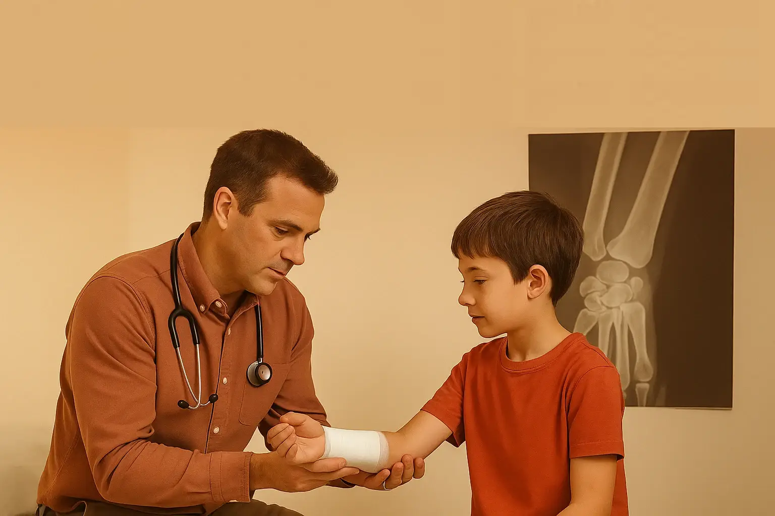
Always order X‑rays for:
If grossly displaced, gently realign (reduce), assess neurovascular status pre- and post-realignment, then immobilise in a padded splint (emedicine.medscape.com, pmc.ncbi.nlm.nih.gov).
For example, humeral supracondylar fractures should be immobilised ~30° from full extension before X‑ray (rch.org.au).
Refer directly if:
Use a systematic ABCS approach (geekymedics.com):
Always get at least two orthogonal views—“one view is no view” (geekymedics.com).
Scaphoid fractures can be occult—initial X‑rays may miss them. If clinically suspected, immobilise in a thumb spica, then review or refer for MRI or CT at 10–14 days ().
Non-surgical cases:
Monitor for red flags:
Q: When should a GP re‑X‑ray after immobilisation?
Routine X‑rays aren’t needed unless symptoms change. One follow‑up image at 3–6 weeks is usually sufficient (emedicine.medscape.com).
Q: Suspected scaphoid fracture, negative X‑ray—what now?
Treat as fracture: immobilise in thumb-spica, review clinically in 10–14 days or arrange advanced imaging (MRI/CT) (northeasthealth.org.au).
Q: What indicates urgent referral?
Open fractures, significant displacement, neuro‑vascular compromise, joint involvement, or suspected compartment syndrome.
Q: How do Ottawa knee rules help?
They guide imaging decisions: age ≥ 55, fibula head/isolated patella tenderness, inability to flex to 90°, weight‑bearing inability—all high‑sensitivity criteria (emedicine.medscape.com, racgp.org.au).
Q: How often to monitor healing?
Typically check at 3 weeks (upper‑limb) or 6 weeks (lower‑limb/femur). More frequent if repositioning was needed or healing seems delayed.
GPs are key to managing many uncomplicated fractures: immobilisation, pain relief, clinical and radiographic follow‑up, and timely referral when needed. Structured X‑ray review and awareness of red‑flag features ensure optimal patient safety and outcomes. When in doubt, early consultation keeps patients well‑served.